Tadalafil gehört zur Gruppe der PDE5-Hemmer und wirkt über eine hochselektive Blockade des Enzyms Phosphodiesterase Typ 5. Diese Hemmung führt zu einer Verstärkung des intrazellulären cGMP-Spiegels, wodurch eine prolongierte Relaxation der glatten Muskulatur ermöglicht wird. Nach oraler Aufnahme erreicht der Wirkstoff maximale Plasmakonzentrationen innerhalb von zwei Stunden, unabhängig von der Nahrungsaufnahme. Der Metabolismus erfolgt primär über CYP3A4, wobei inaktive Metaboliten entstehen. Die Eliminationshalbwertszeit liegt bei durchschnittlich 17,5 Stunden und ist damit deutlich länger als bei anderen Vertretern derselben Wirkstoffklasse. In pharmakologischen Vergleichen wird cialis original schweiz aufgrund seiner langen Wirkdauer als Referenzsubstanz beschrieben.
Bii news
Biomedical Imaging Institute – Newsletter Message from the Director – Prof Geoff Parker Dear Colleague, The new University of Manchester Biomedical Imaging Institute (BII) is uniquely placed to promote and enhance biomedical imaging research within Manchester and
as such will become a centre of excellence on the international stage. The University has considerable expertise in established research areas such as PET, MR, and image
analysis and additional strength in the development of newer technologies such as
electrical impedance tomography (EIT). We are also fortunate to have some of the best available PET and MR imaging facilities located in the Wolfson Molecular
Imaging Centre on the Christie Campus, in the Wellcome Trust Clinical Research Facility on Grafton Street, in the Stopford Building, and in the Translational Imaging
Unit Building at Hope Hospital. These talents and facilities are put to use in answering scientific questions in areas such as neuroscience, oncology,
musculoskeletal disease and cardiovascular disease. The combination of our expertise,
our facilities, and a wide range of preclinical and clinical research application areas puts us in a strong position to compete successfully with the best imaging researchers.
The new Institute is designed to make the most of the University’s imaging capability
The development of new imaging methods The use of advanced imaging methods in scientific clinical and preclinical
Cross-modality integration The integration of pre-clinical and clinical imaging for effective
Act as a unifying structure between University research imaging sites
where we have a concentration of facilities and expertise
Encourage close involvement of Trusts in imaging research (clinicians,
Provide a forum for interaction between basic imaging scientists and
applications researchers to maintain our imaging-related research at the technical leading edge, thereby enabling competitive new findings
Bring through new modalities and methods to biological and clinical
Develop and co-ordinate imaging-related educational activities Provide a focal point for large-scale funding initiatives
If you would like to contribute news articles, please send them to Vicky Catterall 1
Due to the broad-ranging influence of imaging in biomedical research, membership of
the Institute will be an informal affair designed to create an extended community of researchers with overlapping interests. We have attempted to create a circulation list
of individuals who are likely to be interested in our activities but we have undoubtedly overlooked some significant potential contributors. We therefore ask you to circulate
this newsletter widely to colleagues who may be interested and encourage them to get in touch if they wish to be involved.
An official launch event is being organised for 1st May. If, in the interim, you would like to find out more about the BII, would like to contribute to Institute activities, or
would like to comment on the Institute’s formation then please contact me directly ([email protected]) or our coordinator Vicky Catterall
([email protected]). Best wishes,
dical Imaging Symposiu
A monthly symposium series began in Octo
ber 2007 which is designed to provide an
l for anyone who is involved in or consider
s will hopefully leave the symposium knowing
enough to take the first steps in using a gi
ven methodology; they will be told who the
“Independent Component Analysis of functional brain imaging data”
<led by Dr Ingo Schiessl and Dr Hujun Yin>
Venue: Cordingley Lecture Theatre, Humanities Bridgeford Street
The symposium will give a basic introduction to the concepts used in independent
component analysis (ICA). ICA has the ability to reconstruct original signal sources
that are underlying recorded mixtures like brain signals and imaging or biological
artefacts. Without going into mathematical details we will give an overview of the
different approaches available and will demonstrate some results from functional
brain imaging data. We will put a special emphasis on the ability of the method to
detect unexpected components in the results. No special mathematical knowledge is
If you would like to contribute news articles, please send them to Vicky Catterall 2
necessary to follow this symposium which is aimed at researchers in biomedical and
Please email Vicky Catterall if you plan to attend
If you would like to lead a symposium, please contact us - we are currently looking for
speakers for the Summer months. New appointments Dr Oliver Dorn will join the Inverse Problems Group in the School of Mathematics this Summer. He has a wide range of interests in reconstruction in medical imaging
including infra red optical tomography, microwave medical imaging, flourescence tomography, molecular imaging and electromagnetic induction tomography.
He is an international authority on the use of the level set method in inverse
problems, a method especially useful for imaging problems where there are homogeneous regions with smooth boundaries. The method is also used in 2D and 3D
His appointment adds to strength and breadth of the Inverse Problems Group, which
has an international reputation in reconstruction algorithms in electrical impedance tomography. The group also has three PDRAs and three PhD students working on X-
ray tomography. Although this is focussed on applications to security and materials science rather than medical imaging the mathematical techniques are transferable.
Dr Alexander Gerhard recently joined the Clinical Neurosciences group in the School of Translational Medicine and is based at the Wolfson Molecular Imaging
Prof Juri Gelovani has been given a 20% Chair position in Diagnostic Imaging (Cancer Studies Research Group, Cancer & Imaging Sciences). He will develop links
with MD Anderson, his primary employer in Texas, to develop imaging in Oncology.
Prof John Waterton has been given a 20% appointment as Chair in Translational
Imaging (Imaging Sciences Research Group, Cancer & Imaging Sciences). John also
works for AstraZeneca as Chief Scientist in Translational Sciences and has collaborated with the University since 1983. His interests include: imaging
biomarkers in cancer, musculoskeletal and other diseases; the discovery, development and evaluation of such biomarkers; and their translation from the preclinical
If you would like to contribute news articles, please send them to Vicky Catterall 3
New Equipment – Console for 7T Animal Scanner in Stopford Building
Following the award of a BBSRC Equipment Grant before Christmas to Steve Williams (Cancer and Imaging Sciences), David Buckley (Cancer and Imaging
Sciences), Simon Luckman (Faculty of Life Sciences), Stuart Allan (FLS), Mark Boyett (Cardiovascular Medicine) and Ian Stratford (Pharmacy), we have now received
tender responses from suppliers and are in the process of evaluating them. The grant will allow a comprehensive upgrade of the console and bring the small animal facilities
up to state-of-the-art. We hope the new scanner will be up and running by September
of this year. Optical Coherence Tomography at Manchester
There is a growing network of activity in Optical Coherence Tomography (OCT) in
Manchester. A network has been formed, the Manchester OCT Research Network (ManOctNet) which is a joint initiative between the PSI, the medical and dentistry
schools at The University of Manchester, the Manchester Royal Infirmary, and Hope Hospital. In addition to the development and application of bespoke equipment and
techniques for application in the medical and biological fields, the network aims to
increase the awareness of optical methods and capabilities and foster interdisciplinary collaborations.
Optical Coherence Tomography (OCT) uses partially coherent light to illuminate
tissue. It produces sub-surface cross-sectional images with micron level resolution by mixing the backscattered light from internal tissue microstructures with reference
light within an interferometer to construct a real time image. Current technology
allows interrogation of 3-4 mm depth of tissue at a resolution below 10 microns. Doppler OCT (D-OCT) is a variation of OCT allowing measurement of blood flow from
the Doppler shift exhibited by the moving blood cells within the tissue. Advantages over other imaging modalities include:
• tissue images at micron scale in situ in real time • no biopsy or damage to tissue allows repeat imaging at same site • allows non-invasive examination of surface tissues (multiple sites with
reduction in sampling error) and minimally invasive examination using endoscopic/fibre optics
There are a number of OCT activities currently underway, including developing a
dedicated Doppler system for microcirculation studies (funded by the Raynaud’s and
Scleroderma Association), fibre probe development (funded by MIMIT), and OCT imaging in experimental models of peritoneal scarring (with industrial backing), and
investigating skin properties of neo-tropical tree frogs.
If you would like to contribute news articles, please send them to Vicky Catterall 4
In addition the Wellcome Clinical Research Facility has purchased a commercial OCT
system for use in clinical trials and there are a number of clinicians already interested in using the equipment. A number of seminars/workshops will be organized in the near future to introduce the
technology and generate interest from potential users. For more information contact Prof Paul Brenchley
). MIMS Tomography “Anti-Seminar”
The Manchester Institute for Mathematical Sciences hosts an "Anti-seminar" on
problems in Tomography roughly every month. Scientists and Engineers from all Schools and sometimes from collaborating
companies come and present their problems at the meetings, which are called anti-seminars as speakers are encouraged to talk about problems that they have not
solved, rather than a typical seminar talk about what they do know how to do! The audience typically contribute helpful suggestions often leading to research
collaborations. Although the meetings are hosted by mathematics it is as common for people applying
tomographic methods to a completely different scientific to suggest a solution. The physics and application may be quite different but the mathematics (eg the radon
transform) is something they have in common. The Anti-seminar is organized by David Szotten in the School of Mathematics and
Prizes and Awards - 2007
Dr Kate Ward (Senior Scientist, Imaging Sciences Research Group, Cancer & Imaging
Sciences ) and her team won the following prizes and awards last year:
Young Investigator Award, June 2007 (Kate Ward) - 4th International
Children's Bone Health Conference, Montreal, ‘Vitamin D status and muscle
function in post menarchal adolescents’.
Young investigator Award, November 2007 (Rebecca Ashby) – ‘An investigation
of the muscle-bone unit in obese compared to non-obese children’. National
Osteoporosis Society 12th Conference on Osteoporosis, Edinburgh International Conference Centre, Edinburgh,
If you would like to contribute news articles, please send them to Vicky Catterall 5
Young Investigator Award, November 2007 (Kate Ward) – ‘The competing
demands of adolescent pregnancy and skeletal development’. National Osteoporosis Society 12th Conference on Osteoporosis, Edinburgh International
Allied Health Professional Award (Lisa Edwards) – ‘The influence of ageing and
muscle on the peripheral skeleton of young adult male’. National Osteoporosis Society 12th Conference on Osteoporosis, Edinburgh International Conference
Best poster in Trust (Central Manchester) Young Researcher May 2007 (Kate
Ward) – ‘Vitamin D status and muscle function in the post-menarchal girls’.
Major New Grants – Imaging Sciences
Imaging Sciences, Cancer & Imaging Sciences have recently been awarded the following grants, bringing the total Awards figure to £2,374,830 since April 2007:
Sue Astley, CRUK - £58,000 – Extension to the CADET II Breast Screening
Tim Cootes/Chris Taylor, Toyota - £312,000 – Prediction of driving performance
Chris Taylor/Charles Hutchinson – EPSRC/DTI - £292,000 – Automated
analysis of knee cartilage thickness (01/09/07 – 31/08/2010
Geoff Parker/Jo Naish – AstraZeneca – £265,830 - MRI of angiogenesis and
tissue oxygenation (01/003/08 – 28/02/11
Kate Ward – CMMC - £56,000 – Do patients with osteoporosis have increased
suffering from dental disease (01/02/08 – 31/01/10)
Tim Cootes/Chris Taylor – EU - £395,000 – Mobile biometry (01/04/08 –
Alan Jackson/Geoff Parker – AstraZeneca - £400,000 – Purchase of new MRI
Tim Cootes – EPSRC - £310,000 – Modelling and matching for 3D objects for
Medical imagine analysis (01/11/07 – 30/10/10)
Judith Adams/Kate Ward – MRC - £130,000 – Life course pathways to ageing in
Geoff Parker – CRUK - £121,000 – Phase 1 trial of 4_(N-(S-glutathionylacetyl)
Kate Ward – NOS - £35,000 - Changes in bone density, geometry and muscle
funtion in males aged 40 to 80 years old in relation to sex hormones
Fellowships – Imaging Sciences
We would like to congratulate Dr Gerrard Thompson (currently a radiology Walport
Fellow, Imaging Sciences, Cancer & Imaging Sciences) for his award of a Cancer Research UK Clinical Research Training Fellowship for 2008-2011, “Multiparametric
Imaging Biomarkers for Radiotherapy Planning in Glioblastoma Multiforme: Biomarker
If you would like to contribute news articles, please send them to Vicky Catterall 6
Discovery”. He will be supervised by Professors Alan Jackson and Geoff Parker and Dr
Charles Hutchinson.
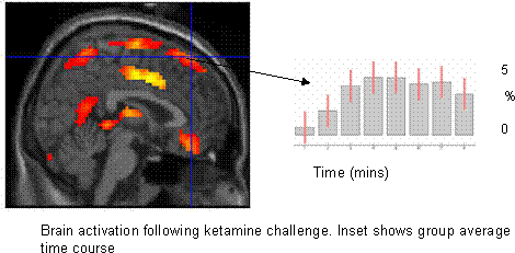
Paper published in top-rated Psychiatry Journal
Bill Deakin and colleagues from the Neuroscience and Psychiatry Unit and ISBE have
published a paper in Archives of General Psychiatry, the top-rated journal for Impact Factor in Psychiatry (>14). The paper is a pharmacological challenge fMRI study of
the effects of ketamine and lamotrigine on regional brain activity and
neuropsychological ratings in normal volunteers. Ketamine is an antagonist at NMDA glutamate receptors and is known to reproduce in normal individuals some of the
symptoms of psychosis. As well as acting as a glutamate receptor antagonist it also promotes glutamate release, so may potentiate glutamate activity at non-NMDA
receptors. By using a secondary challenge with lamotrigine, which blocks all effects of enhanced glutamte release it is possible to dissect the neurochmeical actions of
ketamine. The study showed that ketamine produced a number of regional changes in
brain activity, including areas implicated in schizophrenia pathogenesis. Most of these effects were prevented by pre-treatment with lamotrigine, as were the psychological
effects of ketamine. This proved, for the first time, that ketamine's psychotomimetic actions are mediated by enhanced glutamate release, acting at non-NMDA receptors,
and identified the brain regions underlying this process. This work has given new insights into the neurochemistry and neuroanatomy of schizophrenia and has
supported the glutamate-hyperfunction theory of the disease.
1. Deakin JF, Lees J, McKie S, Hallak JE, Williams SR, Dursun SM. Glutamate
and the neural basis of the subjective effects of ketamine: a pharmaco-magnetic resonance imaging study. Arch Gen Psychiatry 2008;65(2): 154-164
Future Imaging-Related Appointments – Cancer and Imaging Sciences
The School of Cancer and Imaging Sciences have been actively recruiting to two
If you would like to contribute news articles, please send them to Vicky Catterall 7
• A lectureship in neuroimaging will hopefully be filled shortly building on the
• They are in discussion with a senior academic concerning his potentially joining
us for a Chair position in Molecular Imaging in Oncology.
New Projects Approved – Wolfson Molecular Imaging Centre
The WMIC recently approved the following projects:
Gordon Jayson: Biomarkers for the Anti-Angiogenic Anti-VEGF Antibody,
Karl Herholz: The Vascular Contribution to Dementia Karl Herholz: Alzheimer’s disease subtypes: Neuropsychological profile,
Adam McMahon: Development of Quantitative MALDI-MS Imaging
Julian Matthews: Research project into the automatic definition of crystal
Julian Matthews: Research project into the significance of differential detector
sensitivities in the detection of unscattered and scattered events on the HRRT
International Society for Magnetic Resonance Imaging in Manchester (ISMRM)
The University has had 37 papers accepted for ISMRM in Toronto (May 2008). This is
a fantastic achievement for a high level international conference. WMIC granted IMP Licence The MHRA (Medicines and Healthcare products Regulatory Agency) has granted the
Wolfson Molecular Imaging Centre an IMP licence. This is great news as it allows them to manufacture Investigational Medicinal Products on site. Papers published Prof Matt Lambon Ralph’s Neuroscience and Aphasia Research unit (NARU) had its first rTMS paper accepted and published in PNAS – they are the first group to have
If you would like to contribute news articles, please send them to Vicky Catterall 8
G.G. Pobric, E. Jefferies, M.A. Lambon Ralph A selection of recently published papers is listed below:
Journal or book title: Statistics in medicine (In press-published online)
Martínez-Montes E, Cuspineda-Bravo ER, El-Deredy W, Sanchez-Bornot JM, Valdés-Sosa PA,
- The dynamical view of EEG argues that the time locked event related potentials are the result of perturbation of ongoing phase of the EEG. A study, in collaboration with Cuban Neurosciences Centre, on the formal analysis of phase resetting has appeared online in “Statistics in Medicine”.
Journal or book title: Proc Natl Acad Sci U S A Krueger, F.; McCabe, K.; Moll, J.; Kriegeskorte, N.; Zahn, R; Strenziok, M.; Heinecke,
Journal or book title: The new encyclopedia of neuroscience Grafman, J.; Zahn, R; Wassermann, E.
Journal or book title: Annals of the New York Academy of Sciences
Purandare N, Oude Voshaar RC, McCollum C, Jackson A, Burns A.
Journal or book title: Magnetic Resonance Imaging
Journal or book title: Magnetic Resonance Imaging
If you would like to contribute news articles, please send them to Vicky Catterall 9
McGrath, DM; Naish, J; Beatty, PW C; Jackson, A-; Waterton, JC; Taylor, CJ; Parker,
Roberts, C; Parker, GJM; Rose, CJ; Watson, Y; O'Connor, JP B; Stivaros, S; Jackson, A-; Rushton, VE
Kloppel, S.; Draganski, B.; Golding, C.V.; Chu, C.; Nagy, Z.; Cook, P.A.; Hicks, S.L.;
Kennard, C.; Alexander, Daniel C.; Parker, GJM; Tabrizi, S.J.; Frackowiak, R.S.J.
Price, Gary; Cercignani, Mara; Parker, GJM; Altmann, D.R.; Barnes, T.R.E.; Barker, Gareth J.; Joyce, E.M.; Ron, Maria
BII website launched
The Institute’s website has recently been launched and can be found at:
It is still in the process of being developed so you may notice some innaccuracies or
missing information. This will be rectified over the next few months.
If you would like to contribute news articles, please send them to Vicky Catterall 10
EG-Sicherheitsdatenblatt gem. Richtlinie 2001/58/EG Convotherm GmbH Handelsname: CONVOCare K Produkt-Nr.: 3007028 Stand: 07.01.2009 1. Stoff/Zubereitungs- und Firmenbezeichnung Angaben zum Produkt Handelsname: CONVOCare K , Konzentrat zum Aufmischen 1 : 29 mit Wasser Angaben zum Hersteller / Lieferant Adresse: Convotherm Elektrogeräte GmbHTalstraße 35D-82436 Eglfing / G
 Discovery”. He will be supervised by Professors Alan Jackson and Geoff Parker and Dr
Charles Hutchinson.
Discovery”. He will be supervised by Professors Alan Jackson and Geoff Parker and Dr
Charles Hutchinson.