Awh223 2031.2038

The influence of genetic and cardiovascular riskfactors on the CADASIL phenotype Sumeet Singhal,1 Steve Bevan,1 Tom Barrick,1 Philip Rich2 and Hugh S. Markus1 1Clinical Neuroscience, St George’s Hospital Medical Correspondence to: Professor Hugh Markus, Clinical School and 2Neuroradiology, Atkinson Morley Neuroscience, St George’s Hospital Medical School, Neuroscience Centre, St George’s Hospital, London, UK Cranmer Terrace, London SW17 0RE, UKE-mail: [email protected] SummaryThe clinical phenotype in cerebral autosomal dominant dependency or MRI lesion load. There was no evidence arteriopathy with subcortical infarcts and leucoencephalo- of intrafamilial clustering of particular phenotypes.
pathy (CADASIL), an autosomal dominant cerebral Amongst subjects with stroke/transient ischaemic attack, arteriopathy, is variable, but the reasons for this remain smoking at the time of the event was independently asso- uncertain. Possible factors include the mutation site and the ciated with earlier age of onset (P = 0.01). There were no influence of additional modulating factors, which could associations between age of onset or presence of stroke and include both epistatic interactions and interactions with other cardiovascular risk factors, including homocysteine.
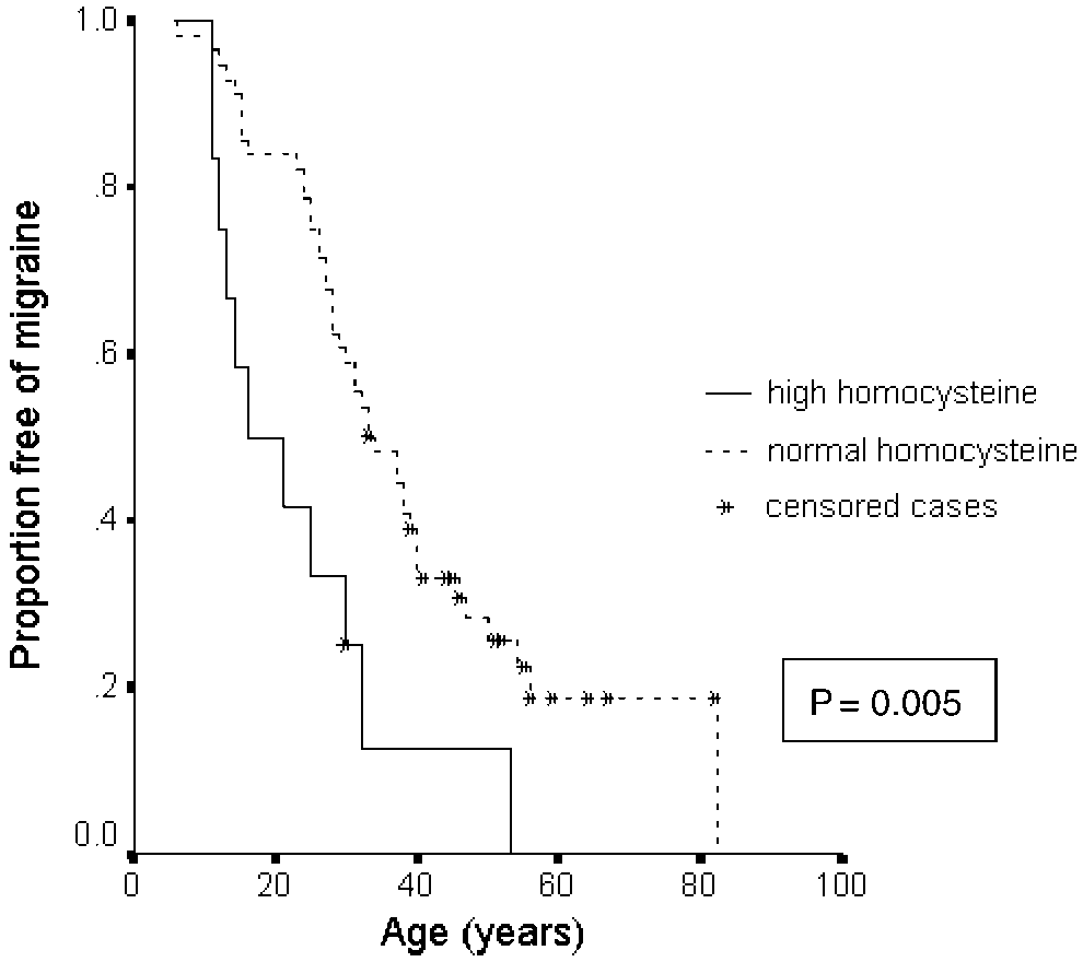
cardiovascular risk factors known to cause sporadic Homocysteine levels were higher in migraineurs [mean small vessel disease. In a large prospectively recruited (SD) 12.8 (5.6) versus 9.8 (3.4) mmol/l, P = 0.02)] and elev- cohort of CADASIL subjects we determined relationships ated homocysteine was independently associated with an between phenotype and mutation site, the apoE genotype earlier age of onset of migraine (P = 0.01). No relationship and cardiovascular risk factors. In addition to clinical fea- was found between MRI lesion volume and risk factors, or tures, disease severity was assessed by MRI lesion volume, between apoE genotype and phenotype. Our results show measured both semiquantitatively (Scheltens scale) and no notch 3 genotype–phenotype correlations. This implies quantitatively. One hundred and twenty-seven CADASIL that modulating factors influence phenotype. Smoking cases from 65 families with 17 different mutations were appears to increase the risk of stroke, while high homocys- studied. Site of mutation was not associated with the teine levels are associated with an increased risk of presence or age of onset of stroke, migraine, dementia, Keywords: CADASIL; genotype–phenotype; homocysteine; risk factors; apolipoprotein E Abbreviations: apoE = apolipoprotein-E; CADASIL = cerebral autosomal dominant arteriopathy with subcortical infarctsand leucoencephalopathy; EGF = epidermal growth factor; MMSE = Mini-Mental State Examination; OR = odds ratio;PCR = polymerase chain reaction; TIA = transient ischaemic attack Received February 17, 2004. Revised April 28, 2004. Accepted April 28, 2004. Advanced Access publication June 30, 2004 IntroductionCerebral autosomal dominant arteriopathy with subcortical cysteine residues. A few splice site mutations (Joutel et al., infarcts and leucoencephalopathy (CADASIL) is an autosomal 2000) and small in-frame deletions (Dichgans et al., 2000) dominant cerebral arteriopathy caused by mutations in the have also been reported, but these also result in an unpaired notch 3 gene (Joutel et al., 1996). Characteristic features number of cysteine residues within the EGF-like domain.
include migraine, recurrent subcortical strokes and subcortical Despite the highly stereotyped nature of the mutations caus- dementia. Mutations are highly stereotyped and result in the ing CADASIL, marked phenotypic variation has been reported gain or loss of cysteine residues in epidermal growth factor (Chabriat et al., 1995; Dichgans et al., 1998). Some individuals (EGF)-like domains in the extracellular portion of the trans- present with stroke in their 20s, while others remain stroke free membrane notch 3 protein. Each EGF-like domain contains into their 60s. The age of onset of the subcortical dementia that three paired cysteine–cysteine bonds. Almost all mutations are frequently develops is variable. Migraine is a common mani- simple missense mutations resulting in an unpaired number of festation, but many patients remain migraine-free throughout Brain Vol. 127 No. 9 # Guarantors of Brain 2004; all rights reserved life. A number of other neurological presentations, including disease progression, particularly the extent of leucoaraiosis, epilepsy and an acute reversible encephalopathy (Schon et al., could be the apolipoprotein-E (apoE) genotype. Presence of 2003), have been reported, but these affect a minority and early the «4 allele predisposes towards Alzheimer’s disease reports suggest they do not seem to segregate within families or (Strittmatter et al., 1993) and has been reported to influence with particular mutations. With the exception of a single report the extent of cerebral damage following a number of suggesting that a novel C455R mutation located in the pre- different brain injuries, including traumatic brain injury and dicted ligand-binding domain may be associated with an earlier subarachnoid haemorrhage (Niskakangas et al., 2001), and age of stroke onset (Arboleda-Velasquez et al., 2002), previous in patients with cerebral amyloid angiopathy (Greenberg studies suggest the site of the notch 3 mutation has little bearing on phenotype. However, there have been limited systematic In this study we examined factors affecting the CADASIL studies examining genotype–phenotype correlations. The lack phenotype in a large cohort of CADASIL individuals recruited of clear genotype–phenotype correlations has led to the sug- from Great Britain. We determined whether the site of the gestion that other factors may modulate the disease process.
notch 3 mutation itself, conventional cardiovascular risk fac- These could include both environmental and other genetic tors, homocysteine and the apoE genotype influenced pheno- factors, but the role of either has not yet been explored.
type. We sought to detect influences both on the extent of CADASIL is characterized by a systemic arteriopathy, visible diffuse cerebral injury, represented by the degree of which specifically affects the cerebral small vessels. Damage leucoaraiosis, and on acute ischaemic injury, presenting as a is particularly severe in smooth muscle cells in the media, stroke or transient ischaemic attack (TIA). We therefore exam- although endothelial changes have also been reported. The ined the relationship between putative risk factors and both most attractive disease hypothesis is that these diffuse changes MRI measures of total leucoaraiosis lesion load, with the result in reduced cerebral perfusion and inability of the cerebral presence and age of onset of stroke/TIA, as well as other vessels to autoregulate. This is supported by imaging studies showing both reduced resting perfusion and impaired vaso- dilatory reserve in response to carbon dioxide (Pfefferkorn et al., 2001) or acetazolamide (Chabriat et al., 2000).
The disease process can result in both acute focal lacunar infarction and more diffuse ischaemic changes, referred to One hundred and twenty-seven CADASIL cases from 65 familieswere studied. Individuals were recruited prospectively as part of a radiologically as leucoaraiosis, and best seen as high signal British CADASIL prevalence study, and all gave informed consent.
on T2-weighted MRI. Lacunar stroke is thought to arise from The study was approved by the South Thames Multi Region Ethics an abrupt disruption of perfusion and/or thrombosis in the Committee. Diagnosis was confirmed by direct sequencing of the perforating end arteries. It could be hypothesized, therefore, notch 3 gene (123 cases), by skin biopsies showing the presence of that any factor either exacerbating the diffuse arteriopathy or granular osmiophilic material in combination with typical neuroima- resulting in an increased tendency to thrombosis might be ging appearances (two cases), and by typical neuroimaging appear- associated with an earlier onset of stroke in patients with ances in a family with biopsy-proven CADASIL (two cases).
The following phenotypic features were recorded: presence and includes ischaemic demyelination, neuronal loss and gliosis age of onset of stroke/TIA; presence and age of onset of migraine; the (Ruchoux and Maurage 1997). It affects the white matter Mini-Mental State Examination (MMSE) score (Folstein et al., 1975); regions supplied by the same perforating vessels affected in the presence of dementia, defined either as a previous diagnosis ofdementia made by a neurologist/psychiatrist, or an MMSE score <23, lacunar stroke. The neuroimaging changes in leucoaraiosis are which has been found to be consistent with DSM-III-R (Diagnostic first seen in regions most distal to the origin of these perforating and Statistical Manual of Mental Disorders) criteria for dementia vessels, i.e. those in which perfusion pressure is lowest. Thus, it (Zaudig, 1992); and a history of acute reversible encephalopathy or is possible that factors resulting either in exacerbation of the primary epilepsy. Functional dependence was measured using the underlying arteriopathy or in increased susceptibility to modified Rankin scale, a six-point scoring system which has been neuronal or other brain tissue injury could increase this validated in the assessment of handicap in stroke patients (van Swieten One explanation for the marked variation in phenotype Using a standard questionnaire and examination, the following could be that vascular risk factors exacerbate the disease pro- cardiovascular risk factors were assessed: hypertension, hypercholes- cess and thus modulate the CADASIL phenotype. These could terolaemia, smoking status, and diabetes mellitus. Hypertension was include both conventional risk factors, such as hypertension defined as an age-adjusted systolic blood pressure >140 mmHg, ordiastolic blood pressure >90 mmHg, or the prescribed use of anti- and diabetes, which are recognized risk factors for non- hypertensive medication. Hypercholesterolaemia was defined as an inherited small vessel disease, and novel risk factors, such age adjusted non-fasting cholesterol >6 mmol/l or the presence of as homocysteine. Elevated homocysteine was reported in a prescribed statin therapy. Smoking was studied in two ways: ever- small study in patients with CADASIL (Flemming et al., smokers at the time of recruitment; and current smokers at the first 2001), and has been shown to be a risk factor for cerebral ischaemic event. In a subgroup of 68 individuals, non-fasting serum small vessel disease, possibly acting via endothelial dysfunc- homocysteine was measured and hyperhomocysteinaemia defined tion (Hassan et al., 2004). An alternative factor influencing results. The logarithm of homocysteine level, the reciprocal of cho- In 112 individuals in whom original MRI scans were available, lesion lesterol level and the square root of MRI lesion load were taken to load was assessed both by the semiquantitative Scheltens scale, and by normalize distributions. For statistical analysis of the relationship of quantitative measurement of lesion volume. The Scheltens scale was phenotype with mutation site, the position of the mutated amino acid in designed for measuring the load and spatial distribution of subcortical the extracellular portion of the notch 3 receptor was determined. The lesions in patients with sporadic vascular white matter disease, and reciprocal of this was used to obtain a normal distribution. We also has been shown to have good intra- and inter-observer reliabilities compared lesion volume and clinical features between patients with (Scheltens et al., 1993). Predefined regions are scored depending first mutations in the predicted ligand binding domain and patients with on size, and then on number of lesions. It was extended to include mutations outside this domain. Patients were categorized as function- scores for the anterior temporal lobe, external capsule and corpus ally independent or dependent if they had Rankin scores of 0–2 or 3–5 callosum, as these regions have all been reported to be frequently respectively. Univariate logistic regression was used to assess correla- affected in CADASIL (Coulthard et al., 2000; Auer et al., 2001; tions between risk factors and categorical outcomes (e.g. presence of O’Sullivan et al., 2001). All scans were scored on the Scheltens stroke/TIA). Linear regression was used for correlations with continu- scale by the same consultant neuroradiologist (P.R.).
ous dependent variables (MRI scores). Multivariate analyses covary- To derive a quantitative measure of lesion load, hard copies of ing for gender and vascular risk factors, in addition to age, were carried T2-weighted MRI scans were digitized and converted to a format out when a statistically significant association was found on univariate suitable for analysis using in-house software (# T. R. Barrick, St analysis. To determine the effect of risk factors on the age of onset of George’s Hospital Medical School, London). White matter lesions, clinical outcomes, Kaplan–Meier analysis was used, followed by Cox the brain circumference and the lateral ventricles were contoured regression to determine the effect of controlling for other potentially using commercially available image analysis software Dispunc (# D. L. Plummer, University College London). Brain parenchymal area was calculated as the difference between brain circumferentialareas and lateral ventricle areas. Infratentorial structures were not included in the analysis, because of incomplete acquisition onsome scans. The most inferior slice for analysis was designated as Clinical characteristics and risk factor profile the first slice to show bilateral anterior temporal lobes as distinct Eight cases identified through family screening were asymp- structures. To account for differences in brain size between subjects, tomatic. They had a mean age of 31 (range 20–45) years, and total lesion load was expressed as a percentage of brain parenchyma.
four (50%) were female. The 119 symptomatic patients had a All scans were analysed by the same person (S. S.). Intra-observer mean age of 48 (range 21–82) years, and 72 (61%) were female.
reproducibility was determined on a random sample of 10 scans. The At recruitment, stroke/TIA had occurred in 71 symptomatic mean of the percentage difference in lesion volume between repeat patients (60%), migraine in 92 (77%), dementia in 22 (19%), lesion load estimation by the same observer was 8.74%. To determine epilepsy in 13 (11%), and acute reversible encephalopathy in within-subjects variation, the mean of the square of SDs of the twoscores for each scan was calculated (Bland, 2000). The within- subjects SD, derived from the square root of this value, was 0.65% Frequencies of cardiovascular risk factors were as follows: [95% confidence interval (CI) À1.15% to þ 2.45%].
hypertension, 24/120 (20%); hypercholesterolaemia, 53/119(45%); current and ex-smoker, 67/126 (52%); diabetes mellitus, 5/127 (4%); hyperhomocysteinaemia, 12/68 (9%).
Mean (SD) cholesterol (n = 115) was 5.6 (1.4) mmol/l, and DNA was extracted from leucocytes using a commercial kit (Nucleon,Amersham Bioscience, Uppsala, Sweden), and polymerase chain mean (SD) homocysteine (n = 68) was 12.2 (5.3) mmol/l.
reaction (PCR) amplification of notch 3 exons was performed. Primersused have been previously described (Markus et al., 2002). Notch 3mutations were sequenced directly with an ABI 377 automated sequencer. Analysis of apoE subtypes was undertaken using pre- viously published methods (Hixson and Vernier, 1990). Genomic The distribution of notch 3 mutations is shown in Table 1. All DNA was amplified by PCR in 10 ml of 75 mmol/l Tris–HCl (pH were simple missense mutations resulting in the loss or gain of a 9), 0.01% (v/v) Tween, 1.5 mmol/l MgCl2, 0.25 U Taq polymerase cysteine residue in one of the EGF-like repeats. After covary- (Abgene, Epsom, UK), 20 mmol/l (NH4)2SO4, 0.1% (w/v) BSA, 0.2 ing for age, site of mutation was not associated with stroke/TIA mmol/l each dNTP and each primer at 5 ng/ml. Thirty cycles of PCR [odds ratio (OR) (95% CI) 1.118 (0.926–1.350), P = 0.25], were performed at 94C for 30 s, 55C for 30 s and 72C for 1 min witha final extension step of 72C for 10 min. Products were then digested migraine [OR 0.985 (0.817–1.188), P = 0.88], dementia with 10 U of restriction endonuclease HhaI (NEB, Hitchin UK) at [OR 0.782 (0.602–1.016), P = 0.07], acute encephalopathy 37C overnight and analysed by 2% low melting point agarose gel [OR 0.938 (0.724–1.215), P = 0.63], primary epilepsy [OR electrophoresis (Abgene). Genotype was determined by comparison 1.008 (0.721–1.409), P = 0.96] or Rankin dependency of the digest pattern to known reference genotypes.
[OR 0.811 (0.623–1.057), P = 0.12]. There was no evidenceof intrafamilial clustering of any particular phenotype, although power to detect this was limited by the small number All dependent variables were analysed with age as a covariate. In of affected individuals recruited in most families.
addition, continuous, normally distributed, independent variables Site of mutation was not associated with quantitative MRI were age-adjusted, and these values were used in all analyses and lesion load [B regression coefficient (95%CI) À0.0003 (À0.075–0.074), P = 0.99] or with total Scheltens score versus 6.56 (4.45) %, P = 0.093]; individual values are [B regression coefficient À0.625 (À1.805–0.554), P = 0.38].
indicated in Fig. 1. Age-adjusted Scheltens score was also Figure 1 shows the quantitative lesion load distribution plotted slightly higher [61.81 (4.38) versus 53.55 (13.39), P = 0.058].
against age, stratified by affected exon.
Three cases, from a single family, had mutations (C440G) in the predicted ligand-binding domain. The individuals were Relationship between vascular risk factors and aged 67, 56 and 37 years and presented with dementia at 66 years, migraine at 14 years and depression at 23 years; none had experienced stroke or TIA. There was a non-significant trend to Stroke/TIA. A history of stroke/TIA was positively associated higher age-adjusted lesion volume in cases with this mutation with age [OR 1.077 (1.040–1.116) per year, P = 0.0003]. After than in cases with other mutations [mean (SD) 9.27 (SD 2.32)% covarying for age, current smoking was associated with anincreased risk of stroke/TIA [OR 2.280 (0.996–5.222), P = Table 1 Mutation spectrum for the 64 families and 0.05]. Following multivariate analysis, covarying for age, gen- 123 individuals where a mutation was identified der, hypertension, hypercholesterolaemia and diabetes, the association was no longer significant [OR 1.961 (0.812– 4.739), P = 0.14]. After covarying for age, there were no asso- ciations between stroke/TIA and gender, hypertension, hypercholesterolaemia, diabetes, ever smoking, serum choles- terol or serum homocysteine (Table 2).
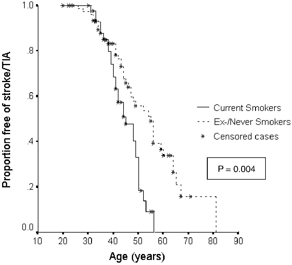
Amongst subjects with stroke/TIA, current smoking at the time of the event was associated with earlier onset of event (P = 0.004, Fig. 2). Following adjustment for gender, hyper- tension, hypercholesterolaemia and diabetes, the association with smoking remained (P = 0.01). On Cox regression there were no associations between age of onset of stroke/TIA and hypertension [OR 0.822 (0.470–1.439), P = 0.49], hypercho- lesterolaemia [OR 0.787 (0.482–1.285), P = 0.34], diabetes [OR 0.847 (0.205–3.507), P = 0.82], serum cholesterol [OR 1.015 (0.958–1.076), per 100/mmol/l, P = 0.61] or serum homo- cysteine [OR 1.002 (0.978–1.026), per 100 Â logmmol/l, P = 0.90].
Migraine. Homocysteine level was associated with age of All amino acid changes involved gain or loss of a cysteine (C) onset of migraine [OR 1.024 (1.004–1.044), P = 0.02]. This remained after controlling for age, gender and all vascular risk Fig. 1 Quantitative lesion load and age, stratified by affected exon. The three exon 8 mutations are in the ligand-binding domain.
Table 2 Association between vascular risk factors or apoE4 allele and presence of clinical features Odds ratios and 95% confidence intervals are given. *P = 0.02; **P = 0.03.
Fig. 3 Kaplan–Meier plot showing the difference in ages of onset Fig. 2 Kaplan–Meier plot showing the difference in ages of onset of migraine between individuals with high (>15 mmol/l) and of stroke/TIA stratified according to smoking status at the time of factors [OR 1.027 (1.006–1.048), P = 0.01]. A Kaplan–Meier covarying for age, there was also a relationship with male plot for migraine age of onset, comparing patients with hyper- gender [OR 3.163 (1.130–8.854), P = 0.03]. There were no homocysteinaemia with normal homocysteine levels, is shown associations with hypertension, hypercholesterolaemia, dia- in Fig. 3. Amongst the 68 subjects in whom homocysteine betes, ever smoking, cholesterol or homocysteine (Table 2).
levels were performed, age adjusted levels were higher in Disability. Rankin score was associated with age at disability migraineurs compared with non-migraineurs [mean (SD) assessment [OR 1.092 (1.044–1.143), P = 0.0001]. After cov- homocysteine 12.8 (5.6) and 9.8 (3.4) mmol/l respectively, arying for age, Rankin dependency was not associated with P = 0.02 (t test on log-transformed data)].
gender, hypertension, hypercholesterolaemia, ever smoking, A history of migraine was not associated with age [OR serum cholesterol or homocysteine (Table 2).
1.001 (0.970–1.032), P = 0.98]. After covarying for age, ahistory of migraine was associated with homocysteinelevel [OR 1.052 (1.008–1.099) per 100 Â log mmol/l, P = 0.02]. After covarying for age, gender and all vascular risk Quantitative MRI lesion load positively correlated with age factors, the association was no longer significant [OR 1.047 [B regression coefficient 0.049 (0.035–0.062), P < 0.00001].
(0.997–1.099), P = 0.066]. A history of migraine was not asso- After covarying for age, lesion load was not associated with ciated with gender, hypertension, hypercholesterolaemia, gender, hypertension, hypercholesterolaemia, diabetes, ever diabetes, ever smoking or serum cholesterol (Table 2).
smoking, serum cholesterol or homocysteine (Table 3).
Scheltens score positively correlated with age [B regression Dementia. A diagnosis of dementia was positively associated coefficient 0.916 (0.709–1.123), P < 0.00001]. After covarying with age [OR 1.082 (1.035–1.130), P = 0.0004]. After for age, there was no association between Scheltens score and Table 3 Relationships of vascular risk factors and apoE4 allele with MRI lesion load,measured quantitatively and using the semiquantitative Scheltens scale Serum homocysteine per 100 Â log mmmol/l B regression coefficients and 95% confidence intervals are shown. No associations were significant.
gender, hypertension, hypercholesterolaemia, ever smoking, mutational hotspot EGF-repeats 2–5 retained the ability to bind serum cholesterol or homocysteine (Table 3).
Jagged 1 and were associated with apparently normal levels ofsignalling activity. In our study there were only three patientsfrom one family with a mutation in this domain. There was a trend to increased age-adjusted lesion volume, measured both quantitatively and with the Scheltens scale, but the numbers are Amongst the 117 individuals in whom successful genotyping too small for firm conclusions. However, such ligand-domain was performed, the apoE genotype frequencies were: e2/e3 mutations account for only a small minority of CADASIL 7.7%, e2/e4 1.7%, e3/e3 76.1%, e3/e4 13.7%, e4/e4 0.9%.
mutations and could not account for most of the phenotypic After covarying for age, there was no association between the presence of at least one e4 allele and dementia, stroke/ Conventional cardiovascular risk factors, such as hyperten- TIA or Rankin dependency (Table 2). There was no association sion and smoking, which are associated with an increased risk between possession of an e4 allele and quantitative MRI lesion of sporadic small vessel disease, could potentially exacerbate load or Scheltens score (Table 3), and mean MRI scores did not the damage to the small cerebral vessels caused by the notch 3 significantly differ for subjects with (n = 17) or without (n = 85) mutation. Such a gene–environment interaction could manifest an e4 allele (lesion load, 6.14 versus 7.25%, P = 0.163; Schel- itself as an increased tendency to acute ischaemia (lacunar tens score, 52 versus 54, P = 0.370).
stroke) and/or more extensive chronic white matter damageseen as leucoaraiosis. Therefore, we examined associationsbetween risk factors and both clinical phenotype and age of onset of stroke, and the extent of leucoaraiosis on MRI. We Our results, from a large prospectively recruited series of used two estimates of MRI lesion volume. First, we used the CADASIL patients from a single country, found no relation- Scheltens score, a semiquantitative scale specifically designed ship between the mutation site and either clinical phenotype or for evaluation of the extent of white matter ischaemic changes the extent of white matter damage on MRI. Furthermore, there (Scheltens et al., 1993). Secondly, we measured lesion volume was no evidence of clustering of particular phenotypes within from MRI scans using an image analysis programme.
families. This is consistent with previous smaller studies We found no association between conventional risk factors, (Dichgans et al., 1998) and suggests that the marked pheno- including hypertension, cholesterol and smoking, and either typic variability seen in the disease is likely to be due to addi- MRI measure of the extent of leucoaraiosis. In contrast, there tional modulating factors. These could include both epistatic was an association between current smoking and age of onset of interactions with other genes and interactions with environ- lacunar stroke, current smokers suffering stroke at an earlier mental risk factors. It has recently been suggested, by study of a age. This suggests that smoking may predispose to episodes of single family, that mutations in the recombination signal bind- acute ischaemia, resulting in occlusion of a perforating artery.
ing protein J Kappa, ligand-binding domain might be asso- Potential mechanisms could include induction of a prothrom- ciated with more severe disease (Arboleda-Velasquez et al., botic state (Hung et al., 1995; Hioki et al., 2001) or impaired 2002). These mutations in EGFR10 and 11 are predicted to be vasomotor function, which has been demonstrated following required for ligand binding by homology to the Drosophila single cigarette smoking in both animals and man (Silvestrini melanogaster notch receptor, and functional studies have et al., 1996; Iida et al., 1998). No other risk factors were reported that such a mutation (C428S) exhibited a significant associated with the presence or age of onset of stroke.
reduction in Jagged1-induced transcriptional activity of a RBP/ Despite the consistent finding that homocysteine levels JK-responsive luciferase reporter, relative to wild-type notch are elevated in patients with sporadic stroke (Hankey and 3, via loss of jagged binding activity (Joutel et al., 2004) In Eikelboom, 2001) and small vessel disease in particular contrast, other mutations (e.g. R90C and C212S) located in the (Bertsch et al., 2001; Hassan et al., 2004), we found no association between homocysteine level and the presence or evidence that conventional cardiovascular risk factors may age of onset of stroke, or the extent of MRI lesion load. In play a modest role in modulating the phenotype in patients contrast, there was a highly significant association between with CADASIL. These results need confirming in prospective elevated levels and migraine. As in other CADASIL cohorts, CADASIL populations. However, they may have important the great majority of cases of migraine were migraine with implications for the management of patients with the disease.
aura. There are a number of potential mechanisms by which The finding emphasizes the importance of smoking cessation.
elevated homocysteine could predispose to this symptom. It Homocysteine levels can be lowered with folic acid and B could act by exacerbating the vascular injury which presum- vitamins (van Guldener and Stehouwer, 2001). Whether this ably leads to both migraine and stroke. However, the lack of will result in a reduction in the frequency of migraine needs to association with stroke or extent of ischaemic damage would be determined in therapeutic trials.
argue against this. Alternatively, there is evidence that homo- Although this is one of the largest series of CADASIL cysteine increases susceptibility to oxidative injury and exci- patients published to date, our findings need replicating in totoxicity, either by activation of the N-methyl-D-aspartate larger populations. Larger studies will require international (NMDA) subtype of glutamate receptors (Lipton et al., collaboration, but similarly sized CADASIL cohorts have 1997), or through DNA damage, p53 activation and subsequent now been identified in a number of countries. Such studies mitochondrial dysfunction (Kruman et al., 2000). All of these will provide further understanding of the reasons for the mechanisms have been suggested as potential mediators in marked phenotypic heterogeneity found in CADASIL, and migraine (Tepper et al., 2001). There is some evidence for may also provide more fundamental information about the an association between migraine and homocysteine in the gen- molecular and other processes resulting in brain damage in eral population. Higher frequencies of the C677T polymorph- response to chronic white matter ischaemia.
(MTHFR) gene, which is associated with higher homocysteinelevels, have been found in patients with migraine compared with controls, and particularly in patients with aura (Kowa We wish to thank Dr Roswell Martin for help with clinical data et al., 2000; Kara et al., 2003). Serum homocysteine levels collection and genotyping, and Dr Yabin Dong and Kelly were not, however, measured in these studies, and a recent Gormley for assistance with genotyping. CADASIL case iden- study failed to find elevated homocysteine levels in migrai- tification was assisted by the British Neurological Surveillance neurs compared with controls. (Hering-Hanit et al., 2001) Unit. This work was supported by the Harrison Lectureship The e4 allele of the apoE genotype has been found to modu- from the Atkinson Morley Neuroscience Research Foundation.
late the extent of brain injury damage in a variety of vascularand non-vascular brain injuries, including Alzheimer’s disease(Strittmatter (Niskakangas et al., 2001) and amyloid angiopathy (Greenberg Andersen K, Launer LJ, Dewey ME, Letenneur L, Ott A, Copeland JR, et al.
Gender differences in the incidence of AD and vascular dementia: The et al., 1995). We have hypothesized that it might also result in EURODEM Studies. EURODEM Incidence Research Group. Neurology more extensive brain injury in response to chronic ischaemia in CADASIL patients. However, no association was found Arboleda-Velasquez JF, Lopera F, Lopez E, Frosch MP, Sepulveda-Falla D, between the e4 allele and the extent of leucoaraiosis on Gutierrez JE, et al. C455R notch3 mutation in a Colombian CADASIL MRI, or with any other features of the disease, including kindred with early onset of stroke. Neurology 2002; 59: 277–9.
Auer DP, Putz B, Gossl C, Elbel G, Gasser T, Dichgans M. Differential lesion patterns in CADASIL and sporadic subcortical arteriosclerotic encephalo- There was an unexpected relationship between male gender pathy: MR imaging study with statistical parametric group comparison.
and the presence of dementia. Some reports have shown gender to be to a risk factor for Alzheimer’s dementia. After Bertsch T, Mielke O, Holy S, Zimmer W, Casarin W, Aufenanger J, et al.
controlling for age, women were at greater risk of developing Homocysteine in cerebrovascular disease: an independent risk factor forsubcortical vascular encephalopathy. Clin Chem Lab Med 2001; 39: 721–4.
Alzheimer’s dementia (Andersen et al., 1999; Molero et al., Bland M. An introduction to medical statistics. 3rd ed. Oxford: Oxford Uni- 2001). Data in vascular dementia have been more varied, but a higher risk has been reported in men (Ruitenberg et al., 2001; Chabriat H, Vahedi K, Iba-Zizen MT, Joutel A, Nibbio A, Nagy TG, et al.
Di Carlo et al., 2002). This may reflect the influence of male Clinical spectrum of CADASIL: a study of 7 families. Cerebral autosomal gender as a risk factor for stroke and therefore for vascular dominant arteriopathy with subcortical infarcts and leukoencephalopathy.
Lancet 1995; 346: 934–9.
dementia (Meyer et al., 2000). This is unlikely to be the expla- Chabriat H, Pappata S, Ostergaard L, Clark CA, Pachot-Clouard M, Vahedi K, nation in our population, because stroke/TIA risk was not et al. Cerebral hemodynamics in CADASIL before and after acetazolamide challenge assessed with MRI bolus tracking. Stroke 2000; 31: 1904–12.
In summary, this study confirms the lack of strong genotype– Coulthard A, Blank SC, Bushby K, Kalaria RN, Burn DJ. Distribution of cranial phenotype relationships in CADASIL. It is possible that MRI abnormalities in patients with symptomatic and subclinical CADASIL.
Br J Radiol 2000; 73: 256–65.
weaker associations may emerge when larger populations Di Carlo A, Baldereschi M, Amaducci L, Lepore V, Bracco L, Maggi S, et al.
are recruited. It is the first study to examine the effect of Incidence of dementia, Alzheimer’s disease, and vascular dementia in Italy.
potential gene–environment interactions and provides some The ILSA Study. J Am Geriatr Soc 2002; 50: 41–8.
Dichgans M, Mayer M, Uttner I, Bruning R, Muller-Hocker J, Rungger G, et al.
apoptosis and hypersensitivity to excitotoxicity. J Neurosci 2000; 20: The phenotypic spectrum of CADASIL: clinical findings in 102 cases. Ann Lipton SA, Kim WK, Choi YB, Kumar S, D’Emilia DM, Rayudu PV, et al.
Dichgans M, Ludwig H, Muller-Hocker J, Messerschmidt A, Gasser T. Small Neurotoxicity associated with dual actions of homocysteine at the in-frame deletions and missense mutations in CADASIL: 3D models predict N-methyl-D-aspartate receptor. Proc Natl Acad Sci USA 1997; 94: misfolding of Notch3 EGF-like repeat domains. Eur J Hum Genet 2000; 8: Markus HS, Martin RJ, Simpson MA, Dong YB, Ali N, Crosby AH, et al.
Flemming KD, Nguyen TT, Abu-Lebdeh HS, Parisi JE, Wiebers DO, Sicks JD, Diagnostic strategies in CADASIL. Neurology 2002; 59: 1134–8.
et al. Hyperhomocysteinemia in patients with cerebral autosomal dominant Meyer JS, Rauch GM, Rauch RA, Haque A, Crawford K. Cardiovascular and arteriopathy with subcortical infarcts and leukoencephalopathy (CADA- other risk factors for Alzheimer’s disease and vascular dementia. Ann NY SIL). Mayo Clin Proc 2001; 76: 1213–8.
Folstein MF, Folstein SE, McHugh PR. ‘Mini-mental state’. A practical Molero AE, Pino-Ramirez G, Maestre GE. Modulation by age and method for grading the cognitive state of patients for the clinician. J associated with the apolipoprotein E-epsilon4 allele in Latin Americans: Greenberg SM, Rebeck GW, Vonsattel JP, Gomez-Isla T, Hyman BT. Apo- findings from the Maracaibo Aging Study. Neurosci Lett 2001; 307: lipoprotein E epsilon 4 and cerebral hemorrhage associated with amyloid angiopathy. Ann Neurol 1995; 38: 254–9.
Niskakangas T, Ohman J, Niemela M, Ilveskoski E, Kunnas TA, Karhunen PJ.
Hankey GJ, Eikelboom JW. Homocysteine and stroke. Curr Opin Neurol 2001; Association of apolipoprotein E polymorphism with outcome after aneur- ysmal subarachnoid hemorrhage: a preliminary study. Stroke 2001; 32: Hassan A, Hunt BJ, O’Sullivan M, Bell R, D’Souza R, Jeffery S, et al. Homo- cysteine is a risk factor for cerebral small vessel disease, acting via endothe- O’Sullivan M, Jarosz JM, Martin RJ, Deasy N, Powell JF, Markus HS. MRI lial dysfunction. Brain 2004; 127: 212–9.
hyperintensities of the temporal lobe and external capsule in patients with Hering-Hanit R, Gadoth N, Yavetz A, Gavendo S, Sela B. Is blood homocys- CADASIL. Neurology 2001; 56: 628–34.
teine elevated in migraine? Headache 2001; 41: 779–81.
Pfefferkorn T, von Stuckrad-Barre S, Herzog J, Gasser T, Hamann GF, Hioki H, Aoki N, Kawano K, Homori M, Hasumura Y, Yasumura T, et al.
Dichgans M. Reduced cerebrovascular CO(2) reactivity in CADASIL: a Acute effects of cigarette smoking on platelet-dependent thrombin transcranial Doppler sonography study. Stroke 2001; 32: 17–21.
generation. Eur Heart J 2001; 22: 56–61.
Ruchoux MM, Maurage CA. CADASIL: cerebral autosomal dominant Hixson JE, Vernier DT. Restriction isotyping of human apolipoprotein E by arteriopathy with subcortical infarcts and leukoencephalopathy. J gene amplification and cleavage with HhaI. J Lipid Res 1990; 31: 545–8.
Neuropathol Exp Neurol 1997; 56: 947–64.
Hung J, Lam JY, Lacoste L, Letchacovski G. Cigarette smoking acutely Ruitenberg A, Ott A, van Swieten JC, Hofman A, Breteler MM. Incidence increases platelet thrombus formation in patients with coronary artery of dementia: does gender make a difference? Neurobiol Aging 2001; 22: disease taking aspirin. Circulation 1995; 92: 2432–6.
Iida M, Iida H, Dohi S, Takenaka M, Fujiwara H. Mechanisms underlying Scheltens P, Barkhof F, Leys D, Pruvo JP, Nauta JJ, Vermersch P, et al. A cerebrovascular effects of cigarette smoking in rats in vivo. Stroke 1998; semiquantitative rating scale for the assessment of signal hyper- intensities on magnetic resonance imaging. J Neurol Sci 1993; 114: 7–12.
Joutel A, Corpechot C, Ducros A, Vahedi K, Chabriat H, Mouton P, et al.
Schon F, Martin RJ, Prevett M, Clough C, Enevoldson TP, Markus HS.
Notch3 mutations in CADASIL, a hereditary adult-onset condition causing ‘CADASIL coma’: an underdiagnosed acute encephalopathy. J Neurol stroke and dementia. Nature 1996; 383: 707–10.
Neurosurg Psychiatry 2003; 74: 249–52.
Joutel A, Chabriat H, Vahedi K, Domenga V, Vayssiere C, Ruchoux MM, et al.
Silvestrini M, Troisi E, Matteis M, Cupini LM, Bernardi G. Effect of Splice site mutation causing a seven amino acid Notch3 in-frame deletion in smoking on cerebrovascular reactivity. J Cereb Blood Flow Metab 1996; CADASIL. Neurology 2000; 54: 1874–5.
Joutel A, Monet M, Domenga V, Riant F, Tournier-Lasserve E. Pathogenic Strittmatter WJ, Saunders AM, Schmechel D, Pericak-Vance M, Enghild J, mutations associated with cerebral autosomal dominant arteriopathy with Salvesen GS, et al. Apolipoprotein E: high-avidity binding to beta-amyloid subcortical infarcts and leukoencephalopathy differently affect Jagged1 and increased frequency of type 4 allele in late-onset familial Alzheimer binding and Notch3 activity via the RBP/JK signaling pathway. Am J disease. Proc Natl Acad Sci USA 1993; 90: 1977–81.
Tepper SJ, Rapoport A, Sheftell F. The pathophysiology of migraine. Neuro- Kara I, Sazci A, Ergul E, Kaya G, Kilic G. Association of the C677T and A1298C polymorphisms in the 5,10 methylenetetrahydrofolate reductase van Guldener C, Stehouwer CD. Homocysteine-lowering treatment: an over- gene in patients with migraine risk. Brain Res Mol Brain Res 2003; 111: view. Expert Opin Pharmacother 2001; 2: 1449–60.
van Swieten JC, Koudstaal PJ, Visser MC, Schouten HJ, van Gijn J. Inter- Kowa H, Yasui K, Takeshima T, Urakami K, Sakai F, Nakashima K. The observer agreement for the assessment of handicap in stroke patients.
homozygous C677T mutation in the methylenetetrahydrofolate reductase gene is a genetic risk factor for migraine. Am J Med Genet 2000; 96: 762–4.
Zaudig M. A new systematic method of measurement and diagnosis of ‘mild Kruman II, Culmsee C, Chan SL, Kruman Y, Guo Z, Penix L, et al.
cognitive impairment’ and dementia according to ICD-10 and DSM-III-R Homocysteine elicits a DNA damage response in neurons that promotes criteria. Int Psychogeriatr 1992; 4 Suppl 2: 203–19.

Source: http://www.cadasil.sgul.ac.uk/the-cadasil-clinic/research/the-influence-of-genetic-and-cardiovascular-risk-factors-on-the-cadasil-phenotype.pdf

Endophthalmitis rates after implantation of the intraocular collamer lens: survey of users between 1998 and 2006

Bruce D. Allan, MD, Isabel Argeles-Sabate, DO, Nick Mamalis, MDAn anonymous on-line survey was sent to 234 intraocular Collamer lens (ICL) (Staar Surgical)surgeons in 21 countries to determine how many of their ICL cases had been complicated by endoph-thalmitis between January 1998 and December 2006. A second questionnaire about the infectiondetails and treatment outcome was sent to those who rep
Microsoft word - j_mccandless.doc

Low-Dose Naltrexone (LDN) for Mood Regulation and Immunomodulation in ASD Jaquelyn McCandless, M.D., April 2006 Naltrexone is a medication used as an opiate antagonist for treating opiate drug and alcohol addiction since the 1970s, FDA-approved since 1985 and now available in generic form as well as in the brand name ReVia in 50mg tablets. At regular dosing to treat addiction,

Copyright © 2010-2018 Pharmacy Drugs Pdf